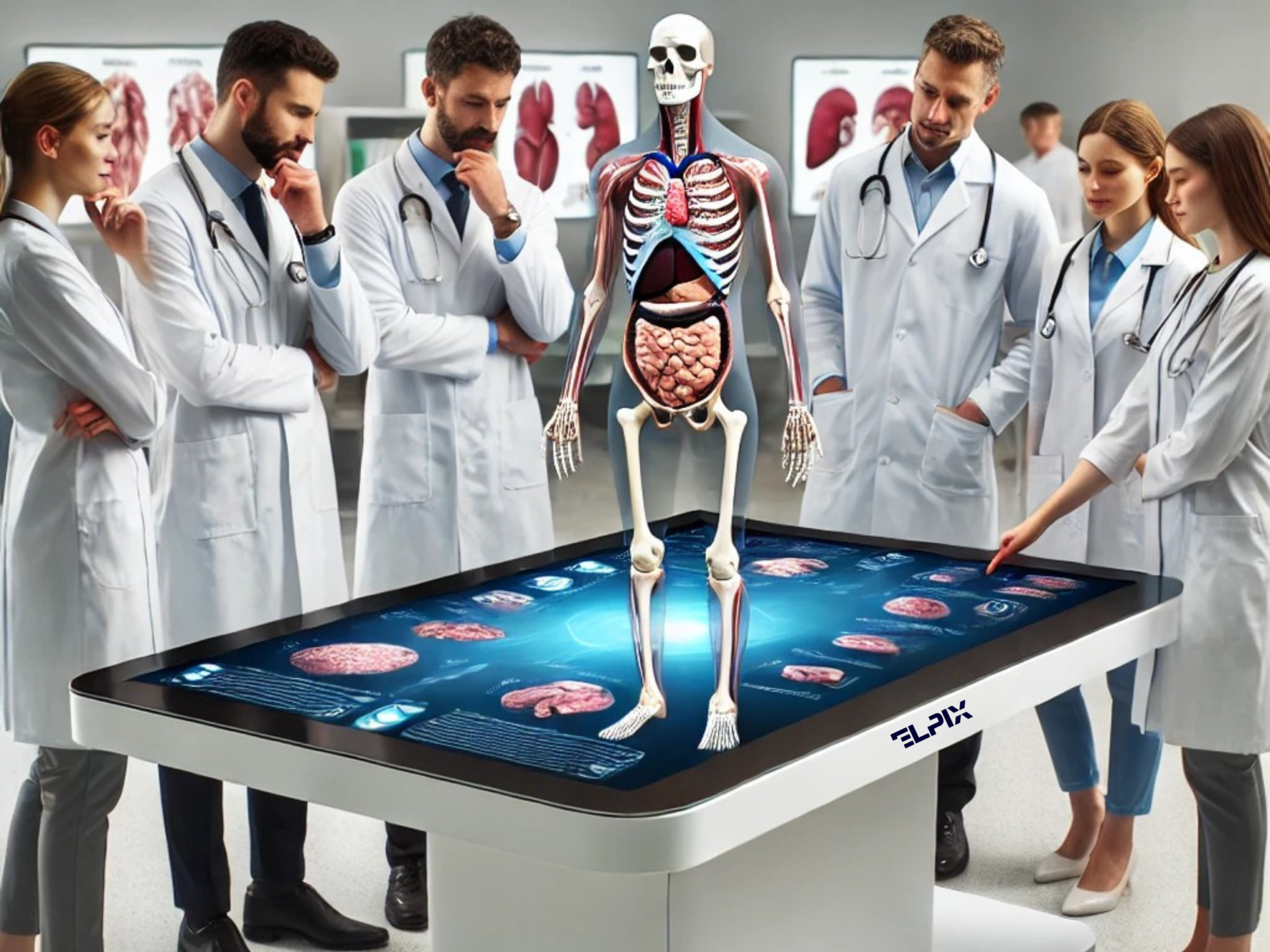
A multimedia anatomical table is not just a gadget for demonstrating human organs. It is a revolution in medical education and medicine in general. Imagine this: instead of traditional textbooks or two-dimensional drawings, you can literally ‘plunge’ into a three-dimensional model of the human body, rotate it, explore it from different angles, and even study the functions of organs and systems in real time.
Multimedia touch tables combine touch screens and 3D technology to allow users to interact with organ models. This is not just an interactive table, but a real medical laboratory that you can hold in your hands. And it’s not a fantasy – such devices are already in active use in universities and hospitals around the world, helping students and doctors stay one step ahead.
The multimedia anatomy table is an innovative device that allows you to interactively study human anatomy in 3D. Using the interactive table, users can interact with three-dimensional models of organs and tissues, change their scale, rotate them and examine them in detail. These tables help to better understand complex anatomical structures, significantly improving the learning process in medical universities and clinics.
The technology allows not only to study human anatomy but also to look at specific pathologies. Imagine how useful it is for students when they are shown what a healthy heart looks like, and then how it changes in case of ischaemia or heart disease. This approach helps them to better understand why the body reacts in a certain way.
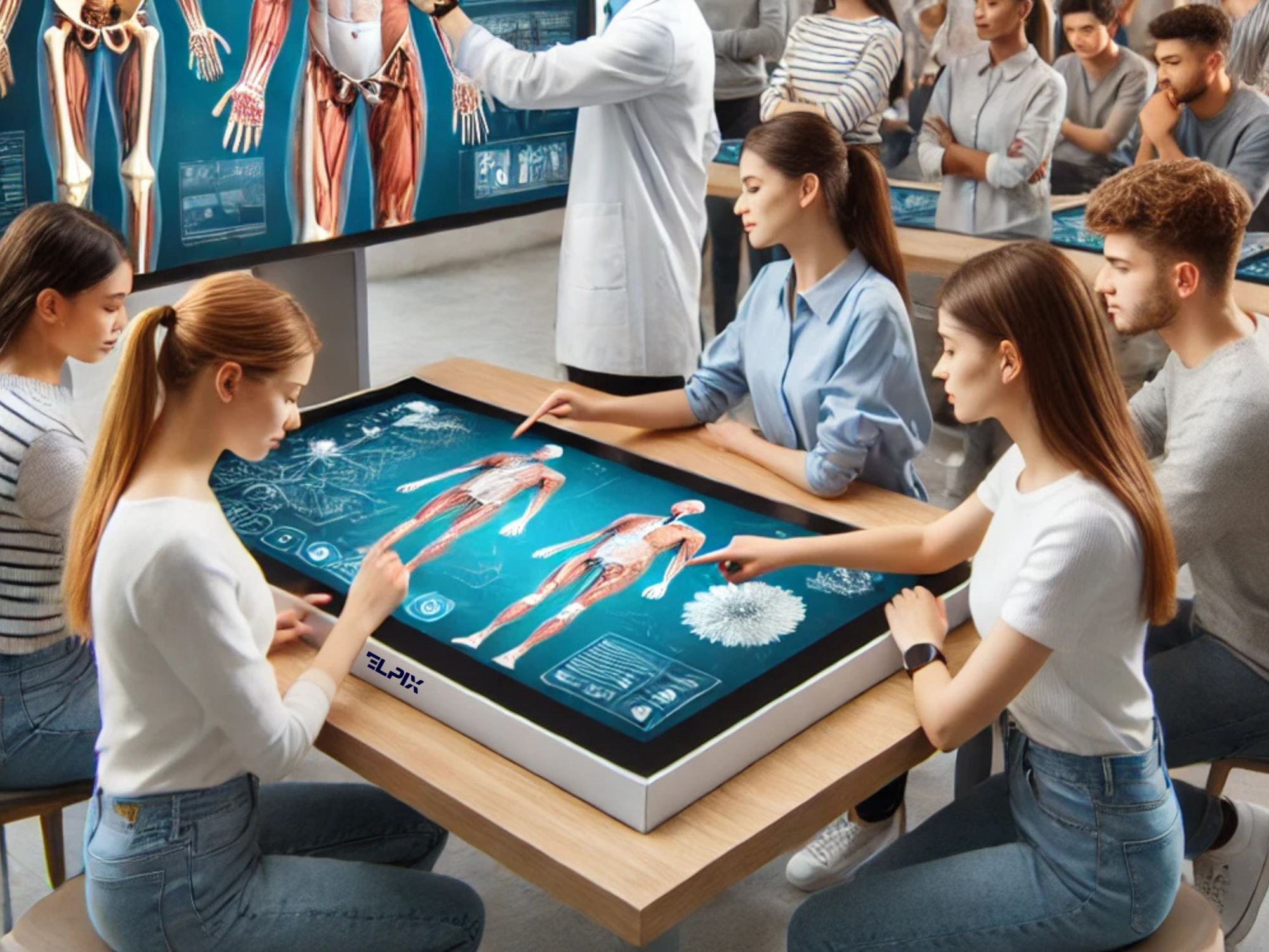
When it comes to learning, especially in medicine, you can’t rely on books alone. The multimedia table is a ‘living’ textbook. It allows students to ‘feel’ anatomy not only at the theoretical level, but also practically. And this is no exaggeration.
Important advantages of such tables are:
Multimedia anatomical tables are not just a trend. It is already a reality that is actively used in many educational institutions and hospitals. For example, the Faculty of Medicine at Uzhhorod National University already has such a table, and it has become an important part of the educational process. Students not only study anatomy, but also have the opportunity to examine various organ pathologies and understand how to treat these diseases in practice. Thanks to this approach, students’ academic performance is significantly improved, and the students themselves note that learning has become much more interesting and effective.
Interactive touchscreen tables are also used in clinics, where doctors can use them not only to perform accurate diagnostics but also to plan surgeries. In the US, for example, such technologies help surgeons perform complex operations with minimal risks to patients.
In addition, the tables are actively used in scientific laboratories to study diseases and organ functions. Such tables help to simulate various situations, studying how the body reacts to specific influences or diseases.
The touchscreen anatomy table is a real boon for medical education and practice. Thanks to its interactivity, 3D models, and the ability to plan operations, this table has become an indispensable tool for students, doctors, and scientists. The use of such tables is already being actively implemented in universities and clinics, and their potential is only growing.
The company Elpix is ready to implement your ideas and implement innovative multimedia solutions that meet the highest standards in medicine and education. If you would like to implement multimedia anatomy tables in your educational institution or medical centre, we will be happy to help you with this, providing not only the equipment but also the technical support for a successful implementation.

